Resume
It has recently been discovered that many animals use information about light polarization as a source of visual contrast. From this type of contrast, animals appear to extract information about shapes and movement that is invisible to us humans. Our main objectives are: to understand which characteristics of light polarization animals use to outline images and how this processing occurs in the nervous system. Using behavioral, electrophysiological, and functional fluorescence imaging techniques, we are trying to understand how polarization information is encoded in the nervous systems of crabs, butterflies, and stomatopod crustaceans. We are also studying in which visual contexts—both biological and non-biological—the use of polarization information is useful for image analysis.
Research
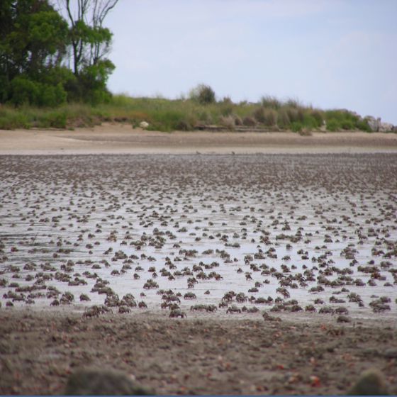
1. Polarized vision in crabs
Using behavioral, electrophysiological, and fluorescence imaging experiments, we investigate which attributes of light polarization animals use to segment the visual scene. (Benjamin Vidal)
2. Polarization image analysis
We study how light polarization information is distributed in the habitats of various animals, with the aim of understanding in which situations analyzing this information can be informative and of developing image analysis algorithms in general. (Gala Minsky)
3. Visual system of butterflies
We recently began studying vision in a fascinating group of animals: butterflies. We are already investigating how color sensitivity is localized within the eye, have started performing electroretinograms, and will soon begin studying their sensitivity to light polarization. (Pablo Riccio and Rodrigo Pampin)
4. Visual system of kissing bugs
Through behavioral, functional morphological, and electrophysiological studies, we are investigating the fundamental aspects of the visual system of the insect vectors that transmit the protozoan responsible for Chagas disease. (Tomás Chialina)
Gallery
Crab Facility 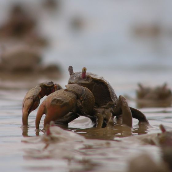
Neohelice crab 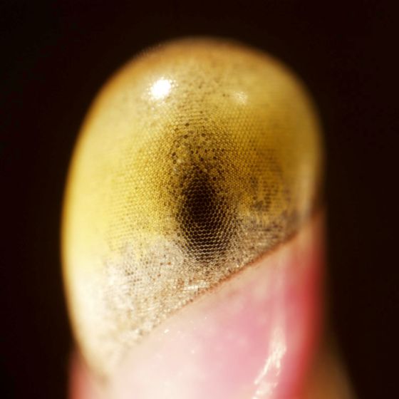
Neohelice Eye 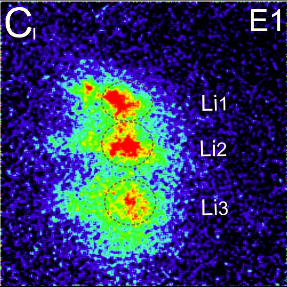
Optic Ganglia Activity
Our Group:
Rodrigo Pampin, MSc
atu.rodrigo@gmail.com
CONICET Doctoral Fellow
MSc in Biological Sciences
Linkedin
Pablo Riccio
.
Undergraduate Thesis Student
.
.
Marcos Sidoruk
.
Undergraduate student in physics.
.
.
Benjamin Vidal, MSc
benjaminlvidal@gmail.com
AGENCIA Doctoral Fellow
MSc in Biological Sciences
Linkedin
Gala Minsky
.
Undergraduate Thesis Student
MSc Student in Biological Sciences
.
Jorge Theodorou
.
Undergraduate student in physics.
.
.
Selected Publications:
- Pérez-Schuster V, Salomón L, Bengochea M, Hermitte G, Berón de Astrada M. Threatening stimuli elicit a sequential cardiac pattern in arthropods. iScience. (2024). Doi: https://doi.org/10.1016/j.isci.2023.108672.
- Basnak M, Pérez-Schuster V, Hermitte G y Berón de Astrada M. Polarized object detection in crabs: a two-channel system. Journal of Experimental Biology. (2018) 25: 221-231.
- Bengochea M y Berón de Astrada M. Organization of columnar inputs in the third optic ganglion of a highly visual crab. Journal of Physiology Paris. (2014). 108:61-70.
- Berón de Astrada M, Bengochea M, Sztarker J, Delorenzi A, Tomsic D. Behaviorally related neural plasticity in the arthropod optic lobes. Current Biology. (2013). 23:1389-98.
- Bengochea M, Berón de Astrada M, Tomsic D, Sztarker J. A crustacean lobula plate: morphology, connections and retinotopic organization. Journal of Comparative Neurology. (2018) 526:109-119. doi: 10.1002/cne
- Berón de Astrada M, Bengochea M, MedanV y Tomsic D. Regionalization in the eye of the grapsid crab Neohelice granulata (=Chasmagnathus granulatus): variation of resolution and facet diameters. J Comp Phys A (2012). 198: 173-180.
- Berón de Astrada M, MedanV y Tomsic D. How visual space maps in the optic neuropils of a crab. Journal of Comparative Neurology. (2012). 519:1631-9.
- Berón de Astrada M, Tuthill JC y Tomsic D. Physiology and morphology of sustaining and dimming neurons of the crab Chasmagnathus granulatus (Brachyura: Grapsidae). J Comp Phy As. (2009) 195: 791-798
Collaborators:
Fernando Locatelli (IFIBYNE, CONICET)
Sebastián Minoli (Dpto BBE, FCEyN-UBA)
Romina Barrozo (Dpto BBE, FCEyN-UBA)
Gabriela Hermitte (Arturo Jauretche National University)